Understanding Bronchial Artery Radiology: The Key to Diagnosing and Treating Hemoptysis
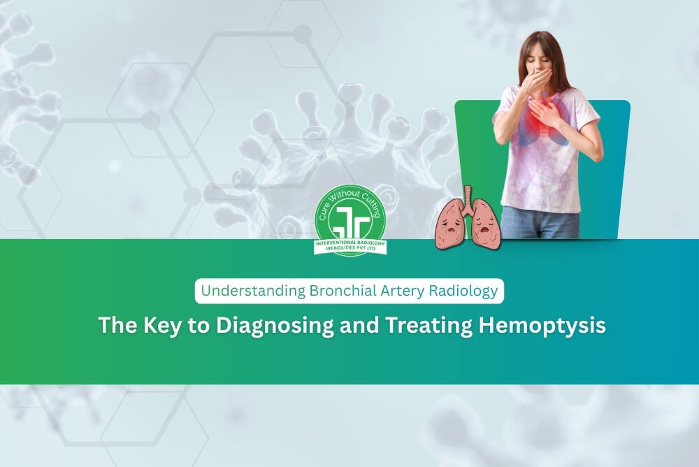
Imagine a sudden onset of bloody coughing and without warning. It's frightening. It's an emergency. And it requires quick answers.
This is precisely the point at which Bronchial Artery Radiology comes in. This highly effective branch of interventional radiology has changed the way doctors identify and manage life-threatening pulmonary ailments. It doesn't matter if you're looking for a patient to better understand the diagnosis, or are simply interested in modern medicine, this book will help you understand everything in a clear, simple manner and with honesty.
So, What Exactly Is Bronchial Artery Radiology?
Let's dissect it.
The lungs require blood to function. The bronchial arterial arteries are the vessels responsible for carrying that oxygenated blood to the airway walls. They're tiny, but essential. If these arteries get large, inflamed, or break due to illness, the effects can be devastating and include massive hemoptysis. This is the process of squeezing out large quantities of blood.
Bronchial Artery Radiology is a specialized field that makes use of advanced imaging techniques including CT angiography as well as the digital subtraction angiography (DSA) -to provide clear images of the arteries. Additionally, it allows doctors to treat the issue without surgery by with a procedure known as embolization of the bronchial artery (BAE).
Imagine this in the following in this way instead of opening the chest cavity, a trained radiologist steers a tiny catheter through the blood vessels in your body, pinpoints the exact cause of bleeding and then seals the area. Precise. Effective. Very minimally invasive.
Why Does This Field Matter So Much?
Here's the real truth: massive hemoptysis could be the death penalty in a lot of cases.Before interventional radiology developed to the point it is at currently, surgeons were required to perform surgery immediately, usually on patients suffering from severe illnesses who were unable to bear the pressure that open surgeries brought. Mortality rates were extremely high. Results were uncertain.
The present day radiology of the bronchial artery has changed the narrative completely. Here's why it is important:
- It pinpoints the problem quickly. Advanced imaging pinpoints the exact blood vessel in minutes, not days.
- The treatment is not making cuts. Bronchial artery embolization is carried out through a tiny cut -- there are no surgical cuts.
- It is lifesaving. Clinical success rates for embolization have been consistently high in the top medical institutions.
- It helps patients return back home earlier. Recovery time is significantly shorter than conventional surgery.
- It decreases the chance. Less trauma to the body translates into less complications after the procedure.
These aren't simply medical data. These are actual changes in real life.
Understanding the Anatomy -- Because It Truly Matters
It is impossible to understand this field without knowing the reasons why it is so difficult.
Bronchial arteries can be wildly variably. They do not have a unique pattern in each person. They may arise from various places along the aorta, differ in size and travel in a variety of ways across the chest. This anatomical complexity is the reason Bronchial Artery Radiology requires deep expertise as well as the latest imaging technology.
In healthy people the arteries function as they should. However, in the case of disease such as bronchiectasis, tuberculosis cystic fibrosis, lung cancer or fungal infections such as the aspergillosis - they turn abnormal. They become thicker. They bend. They become fragile. They eventually bleed.
Without a precise image, finding the vessel that is responsible for the incident is like looking for one thread in an untidy web.
Conditions Where Bronchial Artery Radiology Makes a Real Difference
This area of expertise covers a wide spectrum of serious pulmonary diseases:
1. Tuberculosis (TB)
One of the most frequent causes of hemoptysis is India as well as across the globe. The TB cause an extensive thickening and fragility of the bronchial arteries, which makes them more susceptible to rupture.
2. Bronchiectasis
Chronic lung infections can cause permanent widened airways as well as damaged blood vessels that leak often over the course of time.
3. Lung Cancer
The tumors may erode directly into bronchial vessels, creating sudden and potentially dangerous bleeding.
4. Cystic Fibrosis
Patients are often plagued by repeated hemoptysis episodes over their lifetime which requires multiple embolizations.
5. Aspergillosis and Fungal Infections
Mycetomas, or fungal growths, can slowly degrade vessels' walls, causing moderate but severe bleeding.
6. Pulmonary Arteriovenous Malformations
The abnormal connections between veins and arteries require precise detection and specific radiological treatment.
Each of these ailments are grave. However, each is hopeful due to the precision and accuracy modern radiology has today.
Expert Care That Makes the Difference
In IRFacilities Patients can avail high-quality interventional radiology services that are provided with real clinical expertise and compassion. Under the expert supervision of Dr. Sandeep Sharma, the team takes on each case with meticulous pre-planning, modern technology and a dedication to patient safety.
Since there is something crucial to be aware of that bronchial embolization is not a standard procedure. It requires anatomical expertise and quick decision-making. One of the biggest risk is embolization that occurs accidentally an artery in the spinal cord which could lead to paralysis. This is why choosing an experienced and competent interventional radiologist is not an option. It's essential.
What Does the Future Look Like?
The field is growing rapidly and that's an exciting time for patients.
Technologies such as cone beam CT, 3D rotational angiography as well as AI-powered vessels mapping, are extending the limits of what the bronchial artery radiology can do. These advancements allow for better targeting precision, less risky procedures, and improved long-term results.
In the years ahead we can expect more customized and image-guided strategies to manage pulmonary vascular disease. The aim is simple improved results, quicker recovery, and a better quality of life for each patient.
The Bottom Line
If you or someone close to you is suffering from hemoptysis recurrently or a lung disease that is chronic or any diagnosis that relates to the bronchial vasculature, don't put off getting help.
Bronchial Artery Radiology is not only about imaging. It's about restoring patients' breath, their security and their trust. It's about transforming a terrifying crisis into a manageable, easily treated condition. And it's about having the best expert at your side when it is needed the most.
Seek professional assistance. Make sure you ask the appropriate questions. You can trust a team who is skilled and compassionate -each time.
Ready to Discuss?
Share your symptoms and reports with our team. We help you understand whether you need treatment now or simple lifestyle changes and follow‑up.