Support in Insurance Claim
No-Cost EMI
Without Admission
Short Hospital Stay
4.9 Rating on Google
Benefits

Minimally Invasive

No Visible Scarring

Shorter Recovery Time

No Complications
To Book An Appoinment
What is Aneurysm Coiling?
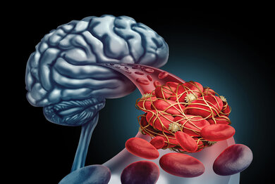
Brain aneurysm coiling specifically treats vulnerable places in brain vessels that sometimes involuntarily expand, resulting in bleeding inside the brain. It is such a procedure in which the doctor threads a long, slender tube into a blood vessel down in the leg and up to the brain- you can't feel anything. Once that's done, it's time for those little metal coils to be packed into the aneurysm, called aneurysm coiling. By these means, blood is discouraged from finding its way into the aneurysm; away from this, an eventual rupture cannot be attained. The method avoids open brain operation, and the brain aneurysm treatment is shorter.

Who are the suitable patients for Aneurysm Coiling?
Eligibility:
This is suitable for patients with small to medium-sized aneurysms, as these may have a small or narrow neck. It is frequently applied to individuals who are at high risk of open brain operation due to the age of the person, medical conditions, and location of the aneurysm. Patients in the emergent stage with recent subarachnoid haemorrhage due to ruptured aneurysm might also benefit from brain aneurysm coiling as an emergency treatment. It is also ideal for some aneurysms located in difficult-to-access brain regions. Case by case, a neurosurgeon or interventional neuroradiologist will assess it using imaging and clinical factors, aiming to make the decision if coiling is the safest and most effective option.
Alternative Treatments:
On the one hand, although aneurysm coiling is an explanation option for many, surgical clipping remains an alternative option where the metal clip is placed on the base of the aneurysm. Clipping could be the preferred method when treating large, broad-necked, or wide-base aneurysms that are difficult to secure with coils. A neuro-interventional radiologist or a neurosurgeon will examine the patient and the aneurysm's characteristics to resolve suitable treatment between coiling and clipping.
Steps of Aneurysm Coiling Procedure
Pre-Procedure Consultation:
Endovascular coiling is deployed inside the aneurysm sac through a minimally invasive approach to treat aneurysms of the brain. The patient would have had either local or general anaesthesia. The catheter would then be inserted through the femoral or radial artery and guided into the brain under imaging. Once the catheter is placed within the sac of the aneurysm, minute platinum coils are released. Clotting induced by endovascular coiling embolization prevents blood from entering the aneurysm, thereby sealing it against rupture. Once an adequate number of coils have been placed, the catheter is withdrawn and the entry site sealed. Afterwards, the patient continues to be managed, including follow-up imaging to confirm that the embolized coil remains patent and that a future aneurysmal bleed does not occur.
Helpful Guidelines for Patients:
Aneurysm coiling for the cerebral artery precedes many moments of preparation that the patient needs to follow. The patient should inform the doctor about all medications taken and allergies. Follow instructions regarding discontinuing blood thinners. No eating or drinking for at least 6 to 8 hours before the intervention. Plan transport to the hospital and aftercare for yourself or a patient. Follow the treatment regarding personal hygiene; for example, shower with antibacterial soap if instructed to do so.


The Aneurysm Coiling Procedure
Step-by-Step Procedure:
Surgery
For the procedure, the patient lies on an angiography table. After the patient is anesthetized, a catheter is placed via the femoral artery or the radial artery, and after it is navigated to the target aneurysm by the use of fluoroscopy, the clot is dissolved, allowing the aneurysm to reorganize, or it is allowed to remain, depending upon the agent used. Coils are then deployed in the aneurysm from inside the catheter, but first, a microcatheter is inserted through the catheter. A platinum coil containing empty space is emptied and placed in the sac of an aneurysm. Inside the aneurysm’s sac, these coils prevent blood from flowing in by tearing the walls of the sac so blood can’t flow through and additionally cause a clot to form that prevents any further blood from entering the aneurysm.
Technology and Tools:
In order for the catheter to reach the aneurysm and deploy the coils very accurately and effectively, fluoroscopy and 3D angiography are used as devices. For wide-necked aneurysms, some patients are included with coiling of the aneurysm with the stents/balloons placed at the same time to aid with keeping the coils in place.
Anesthesia and Comfort:
In some cases, the procedure can be done with local anesthesia with sedation, or in general anesthesia. The vital signs are generally monitored the entire time and the pain is minimal.
Risks and Benefits of Aneurysm Coiling
Advantages:
Aneurysm endovascular coiling has numerous benefits. It is minimally invasive, i.e., no skull is opened, thus reducing the recovery period in comparison to the surgical approach. Most patients end up having shorter hospital admissions in the course of the endovascular treatment and do post-procedure daily activities quickly; therefore, open surgery is often associated with high complication risks.
Disadvantages and Complications:
Coiling of cerebral artery aneurysm, despite being minimally invasive, is still not devoid of risks, as there are chances of infection, blood loss, and even ischemic/hemorrhagic stroke or vasospasm. Also, it is possible, although not probable, that the coils may be misplaced into a non-target artery, resulting in non-target occlusion. Recurrence rates are not uniform, and some patients may need a repeat procedure. Adverse effects may be more common for patients who have complex or wide-necked morphologies because coils can be difficult to keep constant inside the aneurysm.


Recovery After Aneurysm Coiling
Immediate Post-Procedure Care:
After the brain aneurysm treatment, patients are taken to a recovery room, and the duration of their stay depends on various factors. Such factors could include their general condition, for example, whether the patient is healthy or whether there has already been a ruptured aneurysm. The first few effects tend to be slight discomfort situated in the groin (the area where the catheter was inserted) and headaches, which are most often controlled by medications.
Recovery Strategies:
For a couple of weeks, home recovery typically includes rest periods and restrictions on lifting heavy objects, intense exertion, or exercise activities. Medications are often taken to manage pain, and a diet that supports recovery is recommended. Patients are advised to restrict or stop consuming alcoholic drinks and smoking.
Preventive Measures and Prognosis:
Follow-up visits and imaging (such as MRIs or angiograms) are crucial aspects of post-procedural care. Some patients may undergo periodic imaging studies even after treatment to monitor improvement or deterioration over time. In rare cases, if the coil migrates unnecessarily or aneurysm recanalization occurs, further coiling or alternative treatment options might be necessary in the future.


